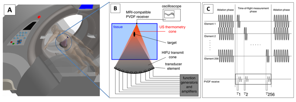
Our innovative research is focused on determining if US tomography-based thermometry in combination with thermal and acoustic modelling can be sufficient to replace MR thermometry and hence devising a system that can perform the same safe and effective uterine fibroid ablations without the need for costly MRI. Our developed system leverages existing HIFU systems by adding external low-cost MRI-compatible US elements (Figure 1). Primarily, these HIFU systems deposit an acoustic thermal dosage but unfortunately omit invaluable intraoperative information. Ultrasound pressure waves going through the ablation zone and propagating to the opposite end carry direct time-of-flight and attenuation information that we intend to record by simply integrating ultrasound elements as shown in Figure 1.
Publications
- Kim Y, Audigier C, Ziegle J, Friebe M, Boctor EM. Ultrasound thermal monitoring with an external ultrasound source for customized bipolar RF ablation shapes. Int J Comput Assist Radiol Surg. 2018; doi: 10.1007/s11548-018-1744-4.