Medical UltraSound Imaging and Intervention Collaboration
Innovation-inspired research
The research objective of the Medical UltraSound Imaging & Intervention Collaboration (MUSiiC) is to characterize fundamental principles at the intersection of robotics, ultrasound physics, bioeffects, signal processing, computer science, and bioengineering, which will enable a new generation of advanced ultrasound imaging technologies capable of providing cost-effective precise interventional guidance and high-quality quantitative and functional diagnostic imaging to a wider sector of people.
The growing availability of ultrasound technology (US) including portable machines has facilitated timely and cost-effective diagnosis in several clinical specialties including trauma, cancer and vascular disease. Ultrasound is an ideal modality due to several factors such as portability, cost, and reduced patient exposure to radiation. Moreover, US is very inexpensive in terms of initial cost and long-term maintenance in comparison to other imaging technologies and produces images at a comparatively rapid rate. It is particularly effective in visualizing blood flow, foreign bodies, fluid effusion, and most large abdominal organs. However, in spite of these many advantages, the capabilities of US remain underutilized because of the following challenges. First, proper US imaging technique requires both static and dynamic loading of an ultrasonographer’s musculature in order to place the probe at an appropriate angle for the acquisition of viable images. In short, sonographers are required to apply high forces for long durations. As a result, 63-91% of ultrasonographers develop musculoskeletal disorders due to the effort required to perform imaging tasks. Second, when diagnosing obese patients with current US imaging systems, it is challenging to achieve high-resolution images for deeply seated targets. In fact, obese patients with high body-mass-index (BMI) may not benefit from many important US exams including vascular exams, abdominal exams, fetal anatomical survey exams, etc.
Goal
Our goal in this research thrust is to create a framework that addresses important needs in today’s diagnostic ultrasound imaging by investigating and developing an economical and practical co-robotic ultrasound imaging platform. Our new co-robotic imaging platform is designed to 1) assist the sonographer by sensing his/her intention and applying needed forces for optimal and safe ultrasound scan, 2) enable high resolution synthetic aperture imaging by providing accurate tracking information to virtually extend the imaging aperture, and 3) provide the accuracy and dexterity needed to enable ultrasound transmission as applied to different quantitative imaging scenarios including prostate and peripheral vascularity.
Relevant publications
Aalamifar F, Cheng A, Kim Y, Hu X, Zhang HK, Guo X, Boctor EM. Robot-assisted automatic ultrasound calibration. Int J Comput Assist Radiol Surg. 2016;11(10):1821-9.
Aalamifar F, Khurana R, Cheng A, Guo X, Iordachita I, Boctor EM. Enabling technologies for robot assisted ultrasound tomography. Int. J Med Robot. 2017;13(1): e1746.
Fang TY, Zhang HK, Finocchi R, Taylor RH, Boctor EM. Force-assisted ultrasound imaging system through dual force sensing and admittance robot control. Int J CARS. 2017; 12(6): 983-991.
Zhang HK, Bottenus N, Cheng A, Guo X, Trahey GE, Boctor EM. Synthetic tracked aperture ultrasound (STrAtUS) imaging: design, simulation, and experimental evaluation. J Med Imaging. 2016;3(2):027001.
Zhang HK, Kim Y, Lin M, Paredes M, Kannan K, Moghekar A, Durr NJ, Boctor EM. Toward dynamic lumbar puncture guidance using needle-based single-element ultrasound imaging. J Med Imaging (Bellingham). 2018;5(2):021224.
Anas EMA, Cheng A, Seifabadi R, Wu Y, Aalamifar F, Wood BJ, Rahmim A, Boctor EM. CNN and back-projection: limited angle ultrasound tomography for speed of sound estimation. Proc. SPIE 10955, Medical Imaging 2019: Ultrasonic Imaging and Tomography, 109550M (10 April 2019).
Seifabadi R, Cheng A, Malik B, Kishimoto S, Wiskin J, Munasinghe J, Negussie AH, Bakhutashvili I, Krishna MC, Choyke P, Pinto P, Rahmim A, Boctor EM, Merino M, Lenox M, Turkbey B, Wood BJ. Correlation of ultrasound tomography to MRI and pathology for the detection of prostate cancer. Proc. SPIE 10955, Medical Imaging 2019: Ultrasonic Imaging and Tomography, 109550C (15 March 2019).
Research Thrust II: Advanced and Multiparametric Ultrasound Imaging
Significance
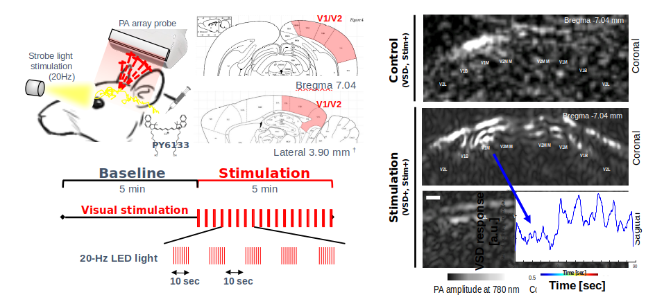
Knowing how the brain works at the cellular and network levels has the potential to greatly improve insight into human disease, and to fundamentally alter our understanding of the human mind. Fundamental issues hinder observation of this extremely complex system, including limited anatomical access to direct observation of neurophysiology and subtle electrophysiological changes. For example, perinatal arterial ischemic stroke is estimated to occur in 1/2300–1/5000 live births and can result in long-term deficits in motor, cognitive, attention, and executive functions and persistent seizures. A great need exists to rapidly diagnose newborns who suffer a focal ischemic stroke, distinguish prenatal vs postnatal stroke, and differentiate perinatal stroke from global hypoxia. Various biophotonic techniques have been proposed for monitoring brain physiology. One of the challenges in the use of neurophotonic applications for brain surveillance has been depth of light penetration. Advanced ultrasound imaging has the potential to address this need by developing new technologies to non-invasively assess neurophysiology and electrophysiological parameters in the brain, and to provide functional and molecular mapping of the cancer environment. Specifically, photoacoustic (PA) imaging is an emerging modality that enables deep tissue, functional imaging. Photoacoustics combines the benefits of both optics and US as it provides the highly specific functional and molecular contrast of photons without suffering resolution degradation as a result of photon scattering in deep tissues.
Goal
Current ultrasound imaging is known to delineate anatomy details, to measure blood flow, and in recent systems to map tissue stiffness. In fact, ultrasound interaction with tissues can reveal even more physiological and functional parameters that are critical to early detection of aggressive cancer, to evaluate brain trauma, or even to provide large scale recording of the brain electrophysiology. We aim in this research thrust to invent, design, and develop algorithms and imaging systems to measure optical properties through photoacoustic (PA) effect, voltage membrane variation, tissue oxygenation, molecular signature, and temperature.
Relevant publications
Zhang HK, Yan P, Kang J, Abou DS, Le HN, Jha AK, Thorek DL, Kang JU, Rahmim A, Wong DF, Boctor EM*, Loew LM*. Listening to membrane potential: photoacoustic voltage-sensitive dye recording. J. Biomed. Opt. 2017;22(4):045006; *corresponding author.
Kang J, Zhang HK, Kadam SD, Fedorko J, Valentine H, Malla AP, Yan P, Harraz M, Kang JU, Rahmim A, Gjedde A, Loew LM, Wong DF, Boctor EM. Transcranial recording of electrophysiological neural activity in the rodent brain in vivo using functional photoacoustic imaging of near-infrared voltage-sensitive dye. Frontiers in Neuroscience 2019; 13:579.
Zhang HK, Chen Y, Kang J, Lisok A, Minn I, Pomper MG*, Boctor EM*. Prostate Specific Membrane Antigen (PSMA)-Targeted Photoacoustic Imaging of Prostate Cancer In Vivo. J Biophotonics. 2018; doi:10.1002/jbio.201800021; *corresponding author.
Anas EMA, Zhang HK, Kang J, Boctor EM. Enabling fast and high-quality LED photoacoustic imaging: a recurrent neural networks-based approach. Biomedical Optics Express 2018; 9(8): 3852-3866.
Kang J, Kadam S, Elmore J, Sullivan B, Valentine H, Malla A, Harraz MM, Kang JU, Loew LM, Baumann M, Grace A, Gjedde A, Boctor EM*, Wong DF*. Transcranial photoacoustic imaging of NMDA-evoked focal circuit dynamics in rat hippocampus. Journal of Neural Engineering 2020; 17: 025001; corresponding author.
Zhang HK, Guo X, Tavakoli B, Boctor EM. Photoacoustic Imaging Paradigm Shift: Towards Using Vendor-Independent Ultrasound Scanners. In: Ourselin S., Joskowicz L., Sabuncu M., Unal G., Wells W. (eds) Medical Image Computing and Computer-Assisted Intervention – MICCAI 2016., vol 9900. pp 585-592.
There has been increasing interest and clinical demand to improve the performance of ultrasound systems in interventional procedures, to reduce the limitations of the ultrasound guidance capability, and to integrate ultrasound into more interventional and surgical applications. A decade ago, under this research thrust, I invented and integrated a miniaturized projector and stereo camera to the ultrasound probe, enabling rapid and non-intrusive probe placement and interventional tool guidance with real-time fusion with prior CT/MRI data. This work has been commercialized by Clear Guide Medical since 2014. Recently, we explored a different approach integrating active ultrasound/photoacoustic systems into the interventional tools. By doing so, a bi-directional ultrasound communication is established between the imaging system and the tool. The tool is actively participating in the image process and forms a closed signal loop in the ultrasound domain. As a result, tracking and imaging in many interventional procedures can be simple, efficient, and reliable.
Goal
This thrust includes multiple research directions. The first direction is focused on developing ultrasonically “smart” interventional tools to facilitate, for example, ablation targeting and monitoring, and catheter guidance without interrupting the current clinical workflow, introducing expensive or intrusive setup, or requiring extensive training. The second direction is to leverage computer vision and deep learning to enable novice users in point of care ultrasound settings. The third direction is a recent effort to investigate and develop “patch” ultrasound technologies.
Relevant publications
Guo X, Kang HJ, Etienne-Cummings R, Boctor EM. Active Ultrasound Pattern Injection System (AUSPIS) for Interventional Tool Guidance. PLoS ONE. 2014;9(10): e104262.
Cheng A, Kang JU, Taylor RH, Boctor EM. Direct Three-dimensional Ultrasound-to-video Registration using Photoacoustic Markers. Journal of Biomedical Optics 18 (6): 066013.
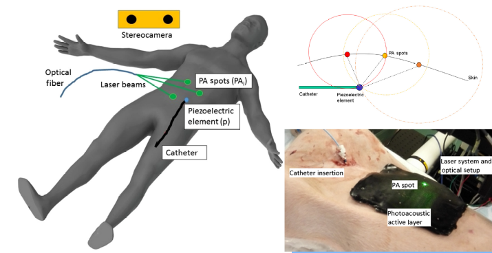
Cheng A, Kim Y, Itsarachaiyot Y, Zhang HK, Weiss CR, Taylor RH, Boctor EM. Photoacoustic-based catheter tracking: simulation, phantom, and in vivo studies. J Med Imaging (Bellingham) 2018 Apr;5(2):021223.
Lediju Bell MA, Guo X, Song DY, Boctor EM. Transurethral light delivery for prostate photoacoustic imaging. J. Biomed. Opt. 2015;20(3): 036002.
Zhang HK, Bell MA, Guo X, Kang HJ, Boctor EM. Synthetic-aperture based photoacoustic re-beamforming (SPARE) approach using beamformed ultrasound data. Biomed Opt Express. 2016;7(8):3056-68.
Kang J, Le HND, Karakus S, Malla AP, Harraz MM, Kang JU, Burnett AL, Boctor EM. Real-time, functional intra-operative localization of rat cavernous nerve network using near-infrared cyanine voltage-sensitive dye imaging. Scientific Report – Nature 2020 (accepted).
Kim Y, Audigier C, Ziegle J, Friebe M, Boctor EM. Ultrasound thermal monitoring with an external ultrasound source for customized bipolar RF ablation shapes. Int J Comput Assist Radiol Surg. 2018; doi: 10.1007/s11548-018-1744-4.
The most successful method of neuromodulation is deep brain stimulation (DBS), which is effective in treating multiple neurological and psychiatric disorders. However, the invasiveness of DBS is associated with many risks due to surgical intervention. Other successful approaches like transcranial direct current stimulation (tDCS) and transcranial magnetic stimulation (TMS) are limited by relatively diffuse spatial resolution (~ 3 cm). On the other hand, focused ultrasound-mediated neuromodulation provides noninvasiveness and high spatial resolution precision (~ 2 mm), providing a solution for current limitations in the field. Cumulative literature over the past 50 years demonstrates that ultrasound modulates neural activity. Ultrasound levels that induce neural activation do not induce thermal or cavitational effects. Our research interest is focused on developing innovative diagnostic and therapeutic tools using ultrasound stimulation bioeffect.
Goal
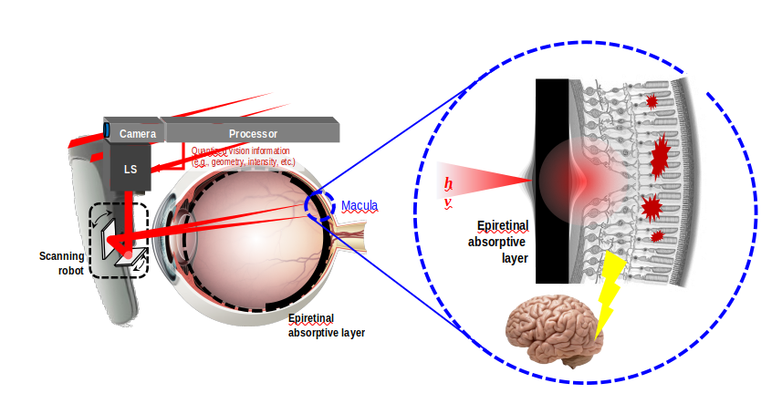
There are more than 12 biological effects induced by modulating ultrasound energy, including hyperthermia, BBB opening, sonoporation, ablation, and neural modulation. Ultrasound as a precise, non-invasive neural activity modulation constitutes a powerful diagnostic and research tool to dissect functional connectivity in the brain and has a plethora of therapeutic applications in psychiatry, neurology, neurosurgery, and ophthalmology. Our research interest is focused on developing innovative diagnostic and therapeutic tools using ultrasound stimulation bioeffect, including early detection of aggressive cancer, accurate quantification of brain damage, and the development of an artificial retina. Additionally, through collaboration with the neuroscience department, we have been investigating the underlying mechanism of action.
Relevant publications
Kang J, Le HND, Zhang HK, Kang JU, Harraz MM, Boctor EM. Initial proof-of-concept of photoacoustic cell stimulation approach: preliminary in vitro study. Proc. SPIE 10052, Optogenetics and Optical Manipulation, 100520U (20 July 2017).
Kang J, Fan Z, Malla A, Harraz M, Spicer J, Gehlbach PL, Boctor EM. An Epiretinal Photoacoustic Stimulation Approach for Retinal Stimulation. Published in Journal Investigative ophthalmology & visual science 2019. Volume 60. Issue 9. Page 4973.
Zhang HK, Kang J, Malla A, Lisok A, Bhujwalla ZM, Pomper MG, Harraz MM, Boctor EM. Bioelectric identification of aggressive prostate cancer using ultrasound cell stimulation. IEEE IUS 2019.
Song H, Ray S, Kang J, Shishikura M, Harraz MM, Boctor EM. Differential Ultrasound Neuromodulatory Responses of Rat Primary Cortical Neurons (PCN) with Oxygen Deprivation in Vitro. IEEE IUS 2020.