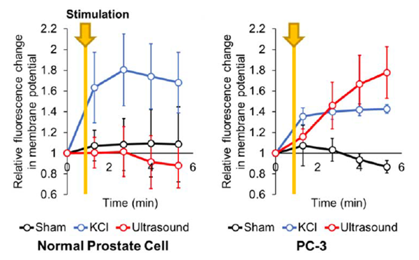
Although many studies suggested that cancerous prostate presents distinct membrane potential, there is currently no active translational research to turn this concept into cancer detection. One of the limitations is the fact that there is no established stimulation protocol and no non-invasive recording of the membrane potential. Ultrasound stimulation has the potential to be the first non-invasive stimulation method that enables clinical translation, and by leveraging our photoacoustic non-invasive voltage membrane variation. We hypothesized that aggressive prostate cancer cells have less polarized potential compared to mild cancer or healthy prostate cells, and are easier to excite under stimulation, since the depolarization threshold can be reached in lower energy stimulation induction. As initial testing, we used two distinct cell types— normal prostate cells, and aggressive cells (PC-3). The preliminary data shows that the PC-3 and normal prostate cells expressed distinct VSD responses when they were exposed to 1-MHz ultrasound neuromodulation (UNM) at 0.53W/cm2 power density for 10 sec from the 1-min time point (Figure 1). Encouragingly, aggressive cell line PC-3 significantly presents fluorescence enhancement compared to that of sham group at the 5-min time point (1.77 ± 0.24 vs. 0.97 ± 0.11, P < 0.01), whereas normal prostate cells present comparable membrane potentials between post- and pre-ultrasound stimulation phases (0.88 ± 0.21 vs. 1.09 ± 0.36). These results indicate that the proposed method can distinguish the selective tissues. Potassium chloride (KCI) serves as the positive control of inducing depolarization. The response upon excessive KCl stimulation is regarded as the positive control validating the operation of utilized slow VSD (i.e., the florescence readout has to be compared at or after the 5-min time point), DiBAC4(3) on both cell types, and also cross validating the excitability of cells. This confirmation leads to the conclusion that indeed two distinct electrophysiological responses were measured from two cell types through US stimulation.
Publications
- Zhang HK, Kang J, Malla A, Lisok A, Bhujwalla ZM, Pomper MG, Harraz MM, Boctor EM. Bioelectric identification of aggressive prostate cancer using ultrasound cell stimulation. IEEE IUS 2019.