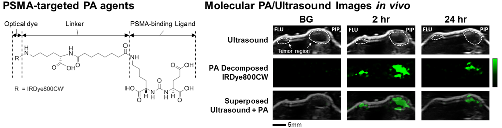
We have developed a molecular PA imaging system capable of visualizing PSMA molecular contrast in vivo. We have designed a series of PA imaging contrast agents composed of three functional parts (Figure 1). The middle component is a linker that tethers the PSMA ligand to bulky optical dyes such as IRDye800CW, ICG, and Alexafluor750, which generated the strongest PA signals among ones we tested. The PA agent using IRDye800CW showed the best sensitivity in vivo (Figure 1). To differentiate PA contrast between blood and dye, the received multi-wavelength data were decomposed to quantify contrast due purely to dye and, by extension, PSMA expression. Results were correlated and validated with the ground truth FL measurements immediately after PA imaging. These studies with our first-generation agents support the potential of PA imaging to dissect subtypes of prostate cancer.
Publications
- Zhang HK, Chen Y, Kang J, Lisok A, Minn I, Pomper MG*, Boctor EM*. Prostate Specific Membrane Antigen (PSMA)-Targeted Photoacoustic Imaging of Prostate Cancer In Vivo. J Biophotonics. 2018; doi:10.1002/jbio.201800021; *corresponding author.