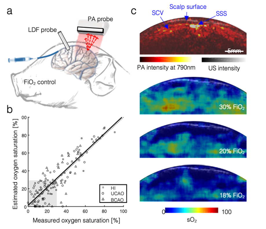
The feasibility of transcranial functional PA imaging of cerebral oxygenation in the piglet brain (5-day old, 2kg) was demonstrated (Figure 1a). Multi-spectral PA data were collected (700-900 nm in 10-nm intervals) while applying various fractional inspired oxygen (FiO2) levels to induce graded hypoxia. The ground-truth sO2 was also measured by direct sampling of venous blood at the superior sagittal sinus (SSS). Figure 1b shows that transcranial functional PA imaging of sO2 is highly correlated to the ground truth. Also, transcranial functional PA sO2 images across the cortical tissue regions were reconstructed (Figure 1c). As a result, evident physiological contrast changes could be visualized in deep piglet brain regions through intact scalp and skull layers. We further validated transcranial functional PA neuroimaging in a piglet model with a photothrombotic stroke. To prepare this work for clinical translation, we have investigated a safer, cost-effective laser source for transcranial PA imaging. Compared to the conventional Nd:YAG OPO laser system, a pulsed light-emitting diode (LED) system is much safer (no need for protective glasses), which makes it preferable for clinical use. Furthermore, we were the first to investigate and report deep learning in enhancing LED-based PA signal and perform automatic detection of hypoxia status.
Publications
- Zhang HK, Yan P, Kang J, Abou DS, Le HN, Jha AK, Thorek DL, Kang JU, Rahmim A, Wong DF, Boctor EM*, Loew LM*. Listening to membrane potential: photoacoustic voltage-sensitive dye recording. J. Biomed. Opt. 2017;22(4):045006; *corresponding author.
- Kang J, Zhang HK, Kadam SD, Fedorko J, Valentine H, Malla AP, Yan P, Harraz M, Kang JU, Rahmim A, Gjedde A, Loew LM, Wong DF, Boctor EM. Transcranial recording of electrophysiological neural activity in the rodent brain in vivo using functional photoacoustic imaging of near-infrared voltage-sensitive dye. Frontiers in Neuroscience 2019; 13:579.
- Kang J, Kadam S, Elmore J, Sullivan B, Valentine H, Malla A, Harraz MM, Kang JU, Loew LM, Baumann M, Grace A, Gjedde A, Boctor EM*, Wong DF*. Transcranial photoacoustic imaging of NMDA-evoked focal circuit dynamics in rat hippocampus. Journal of Neural Engineering 2020; 17: 025001; corresponding author.