Peripheral nerve damage often produces serious problems in human life. By the fact that the peripheral nerve is not protected by rigid organ such as bone, it is at high risk of getting damage during surgery, daily life, and etc. For instance, during nerve sparing radical prostatectomy, peripheral nerves surrounding the prostate gland are easily damaged while removing the entire prostate. This will bring significant post-operative complication to patients such as erectile dysfunction and urinary incontinence. Therefore, preventing the nerve from getting damage is of significant important factor in surgical environment.
The project can be separated into two main parts. These are
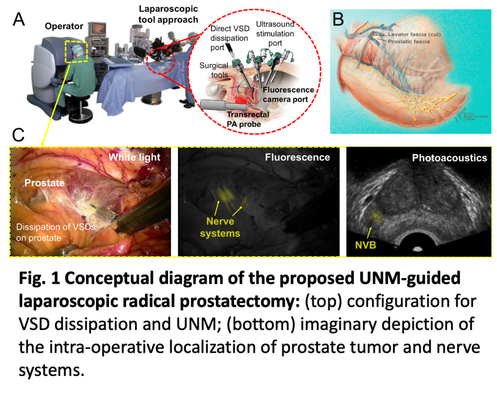
1. Real-time nerve imaging system in da Vinci platform
We implement a real-time system in da Vinci platform, which is capable of monitoring the neuronal activities during a surgery such as prostatectomy. An illustration below indicates the proposed protocol for intra-operative surgical guidance. A transrectal ultrasound (TRUS) transducer will follow the surgical tool tip in order to provide the same imaging plane as the surgical spot. Consequently, photoacoustic signal will be acquired from the laser and the US+PA image will provide the active nerve network in surgeon’s field-of-view while operating.
2. Membrane potential measurement
Quantitative assessments of neuronal activities have been conducted. Voltage-sensitive-dye (VSD) are applied to the nerve fibers, and focused ultrasound is used to stimulate the neurons. The stimulation will cause the cells to depolarize and the resultant membrane potential change will vary the photoacoustic intensity from the VSD. We develop an imaging technique visualize the membrane potential variation by quantitatively measure the photoacoustic signal.
People
Hyunwoo Song [email protected]
…………….
Publication
- Cheng, Alexis, et al. “Ultrasound to video registration using a bi-plane transrectal probe with photoacoustic markers.” Medical Imaging 2016: Image-Guided Procedures, Robotic Interventions, and Modeling. Vol. 9786. International Society for Optics and Photonics, 2016.
- Zhang, Haichong K., et al. “Listening to membrane potential: photoacoustic voltage-sensitive dye recording.” Journal of biomedical optics 22.4 (2017): 045006.
- Kang, Jeeun, et al. “Real-time, functional intra-operative localization of rat cavernous nerve network using near-infrared cyanine voltage-sensitive dye imaging.” Scientific reports 10.1 (2020): 1-10.