Motivation
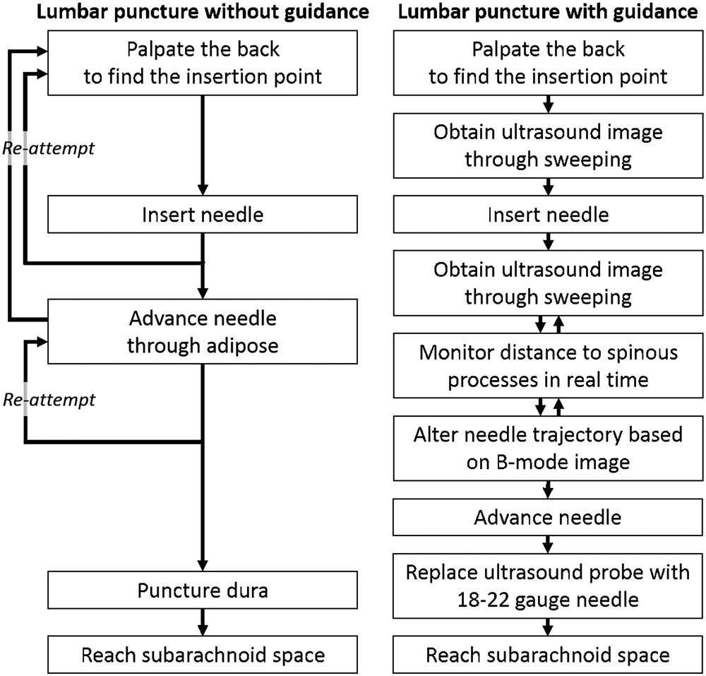
People get lumbar puncture (LP) to diagnose a variety of central nervous system disorders or conditions, including life threatening ones like encephalitis or meningitis. It is usually done blindly: your doctor feels around the spine to find the gap between bones. They guesses the location and angle and inserts the needle. They knows the needle is deep enough by feeling a pop and the CSF should ooze out. However, this procedure is difficult on obese patients. 400,000+ LP are performed annually, but nearly 23% end in failure for various reasons. Failures lead to complications like headaches back pain.
Problem and current solutions
The challenge is in visibility. Some commercial products address this problem by tracking the needle and overlaying on the ultrasound image. Notable examples include eZono eZGuide (magnetic needle tracking) and ClearGuide One (optical needle tracking). They cannot place the ultrasound image in plane with the needle due to physical interference and are prone to needle tracking error due to needle bending.
Our approach
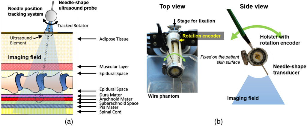
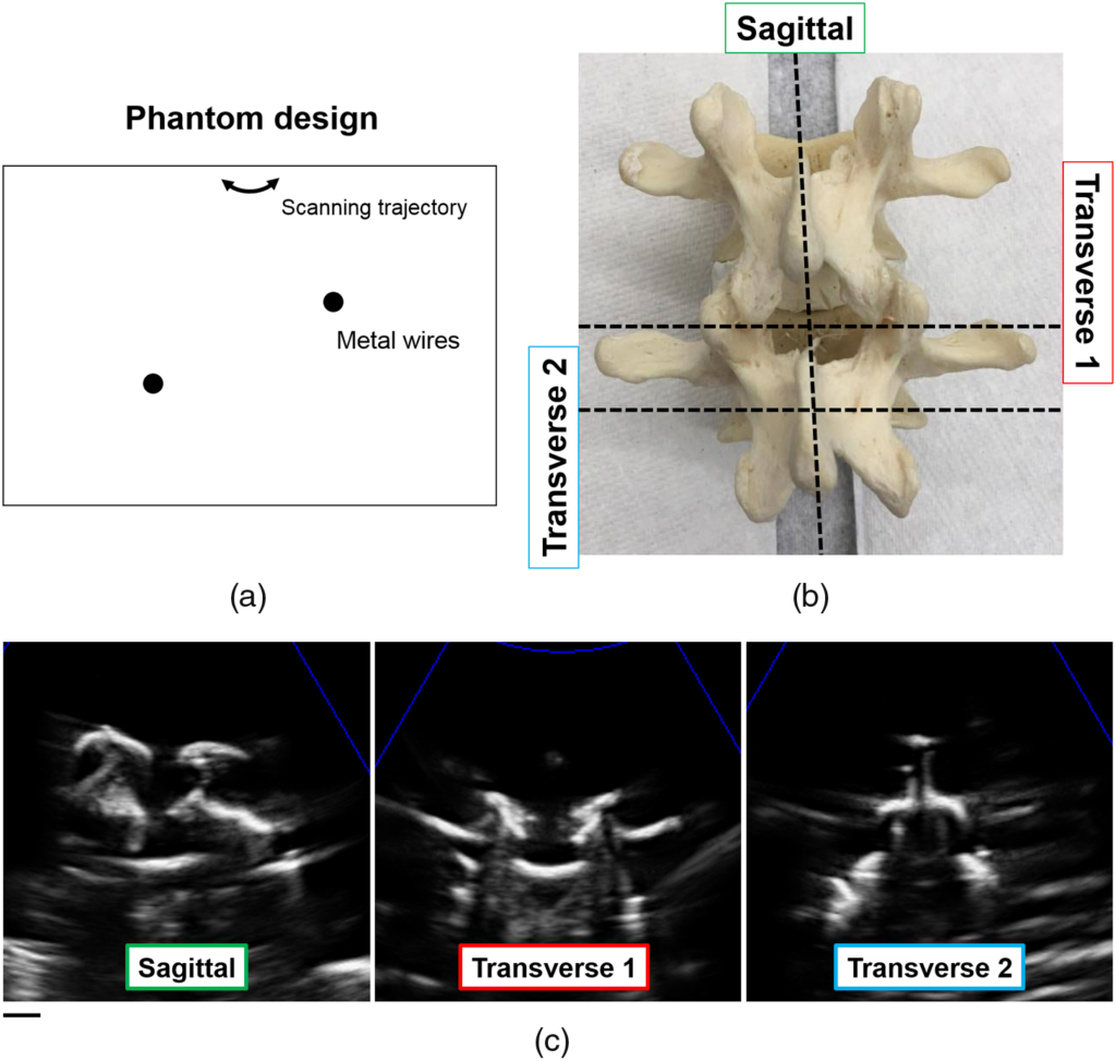
We designed a needle with an ultrasound element in the tip. We sense the angle of the needle with a rotational encoder. When the doctor sweeps the needle, our system builds an image of the tissue below.
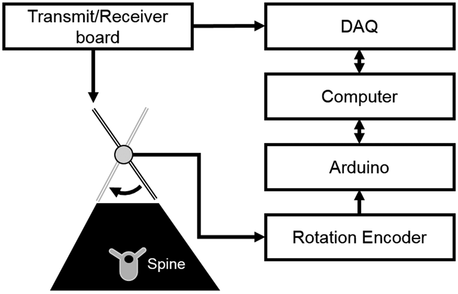

We also actuated the needle with a delta-configuration actuator. It moves the ultrasound needle in 3 translational DOF to build an image.
Publications
- Keshuai Xu, Younsu Kim, Emad M. Boctor, Haichong K. Zhang, “Enabling low-cost point-of-care ultrasound imaging system using single element transducer and delta configuration actuator,” Proc. SPIE 10951, Medical Imaging 2019: Image-Guided Procedures, Robotic Interventions, and Modeling, 109510W (8 March 2019);
- Zhang, H. K., Kim, Y., Moghekar, A., Durr, N. J., and Boctor, E. M., “Single-element needle-based ultrasound imaging of the spine: An in vivo feasibility study,” in [Simulation, Image Processing, and Ultrasound Systems for Assisted Diagnosis and Navigation], 82–89, Springer (2018).
- Zhang, H. K., Kim, Y., Lin, M., Paredes, M., Kannan, K., Moghekar, A., Durr, N. J., and Boctor, E. M., “Toward dynamic lumbar puncture guidance using needle-based single-element ultrasound imaging,” Journal of Medical Imaging 5(2), 021224 (2018).